
Retina Conditions
Retinal Tear and Retinal Detachment
Retinal detachment is a serious condition that can lead to permanent vision loss if not treated quickly.
What is a Retinal Detachment?
A retinal detachment occurs when the retina peels away from the back wall of the eye. A detached retina cannot function properly, and the result is a shadow in your peripheral vision that can spread and cause significant blurring of your sight.
A retinal detachment is a medical emergency. Urgent evaluation and treatment are essential, because the detachment can permanently damage your vision if it is not repaired promptly, often within 24 to 72 hours.

Normal Vision:
Clear visual acuity.

Retinal Tear:
A retinal tear and retinal detachment may cause a curtain or shadow to appear across your field of vision.

Retinal Detachment:
You may see additional floaters and flashes, and images may take on a wavy or watery appearance.
What Causes a Retinal Detachment?
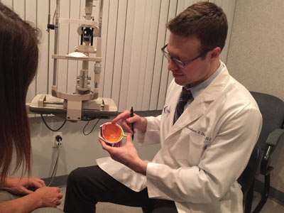
Most retinal detachments begin with age related changes in the vitreous, the gel that fills the eye. As we get older, the vitreous shrinks and liquefies. Normally it moves freely within the eye as the eye moves, without disturbing the retina.
Occasionally, however, the shifting vitreous pulls on the retina hard enough to create a tear or rip. Once a tear is present, fluid from inside the eye can pass through the opening and lift the retina off the back wall of the eye, the way water seeping under wallpaper peels it from the wall. That lifting is the detachment.
Who is at Risk for a Retinal Detachment?
Your risk of retinal detachment is higher if any of the following apply to you:
• Nearsightedness, meaning you need glasses to see far away.
• Previous cataract surgery.
• A history of glaucoma, an eye disease related to eye pressure.
• Prior serious eye injuries.
• A history of a retinal tear or detachment in your other eye.
• A family history of retinal detachment.
• Weak areas in your retina, called lattice degeneration, seen by your eye doctor on previous exams.
What Are the Early Warning Signs of a Retinal Detachment?
The warning signs of a retinal detachment are sudden flashes, new floaters, and a shadow or curtain in your side vision. Without timely treatment, permanent vision loss can result, so call your eye doctor right away if you notice any of the following:
• Flashing lights, such as seeing stars similar to being hit in the eye.
• New floaters or a sudden increase in floaters, which can look like specks of dust, lines, mosquitos, flies, or cobwebs drifting through your vision.
• A shadow or grey curtain appearing in your peripheral (side) vision.
How is a Retinal Detachment Diagnosed?
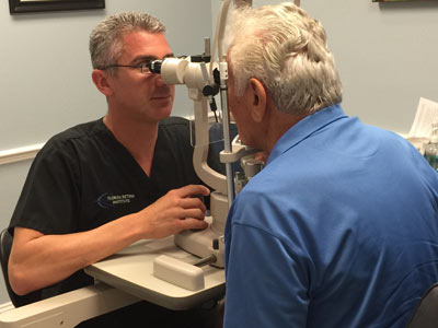
A retinal detachment is diagnosed through a dilated eye exam. Your eye doctor places drops in your eyes to widen the pupils, then performs a slit lamp evaluation, positioning special lenses between the slit lamp and the cornea, or directly on the cornea, to view the deeper structures of the eye, including the optic nerve, the retina, and the drainage angle where fluid exits the eye.
A camera may be attached to the slit lamp to photograph different parts of the eye. This allows your doctor to carefully examine the peripheral retina, which is where most detachments are located.
Dr. Mavrofrides performing a slit lamp examination on a patient.
How is a Retinal Detachment Treated?
A retinal detachment requires surgery, performed either in the office or in the operating room. There are several ways to repair a detached retina:
Pneumatic retinopexy. This procedure can be performed in the office. Your retina specialist places a gas bubble inside the eye, where it acts as a splint that holds the retina in place so it can heal properly. Afterward, you will need to keep your head positioned a specific way for a few days to keep the bubble against the right spot. As the eye heals, the gas bubble is absorbed on its own over a few weeks.
Vitrectomy surgery. This procedure is performed in the operating room. Your retina specialist removes the vitreous gel from the eye, eliminating the pulling on the retina. A full gas or oil bubble is then placed in the eye to support the retina like a cast while it heals. A gas bubble dissolves over a few weeks; if an oil bubble is needed, your surgeon removes it in a second procedure a few months later.
An important safety note: while you have a gas bubble in your eye, you cannot fly in an airplane. Altitude changes cause the gas to expand, which raises the eye pressure and can result in permanent vision loss.
Scleral buckle. A soft silicone band is attached to the outside of the eyeball, behind the eyelids, where it gently indents the eye inward. This relieves the pulling of the vitreous gel on the retina.
What Are the Risks of Retinal Detachment Surgery?
Every surgery carries some risk, but the comparison here is stark: without treatment, a retinal detachment leads to permanent loss of vision. Possible surgical risks include:
• Infection in the eye.
• Bleeding in the eye.
• Increased eye pressure.
• Clouding of the lens inside the eye, known as a cataract.
• Scar tissue formation that can detach the retina again.
• The chance that the retina does not reattach properly.
• The need for additional surgery.
Your vitreoretinal surgeon will discuss the risks and benefits of surgery with you before proceeding.
What Should I Expect After Retinal Detachment Surgery?
Recovery from retinal detachment surgery takes patience, and knowing what is normal helps. Here is what to expect:
• You may have some pain for a few hours after surgery, and you will be given pain medicine to keep you comfortable.
• You will need to rest and limit your activity for a few weeks. Your retina specialist will tell you when you can exercise, drive, or resume other activities.
• You will wear an eye patch after surgery. Keep it on as long as your doctor instructs.
• If a bubble was placed in your eye, you will need to hold your head in a specific position for about 1 to 2 weeks. Your retina specialist will show you the position, and following these directions closely is essential for proper healing.
• You may see floaters and flashing lights for a few weeks after surgery, and you may notice the bubble itself. Over time the bubble shrinks and may break into smaller bubbles before disappearing.
• Your sight should begin improving about four weeks after surgery, though your vision may keep changing for months. In some cases, complete healing can take a year or more.
How Quickly Does a Retinal Detachment Need to Be Treated?
Immediately. A detached retina lifts away from the back of the eye, and every hour it remains detached puts your sight at greater risk. The warning signs are flashing lights, many new floaters, a shadow in your side vision, or a grey curtain covering part of your field of vision. If you experience any of these signs, call an ophthalmologist right away. A detached retina must be treated urgently to preserve your vision.
If you are experiencing flashes, a sudden increase in floaters, or a shadow or curtain in your vision, do not wait. Please contact Florida Retina Institute or call our office immediately to speak with a member of our team. With 21 locations throughout Florida and Georgia, urgent retina care is always close to home.