About Us / Blog and News / NGENUITY 3D Digital Retina Surgery
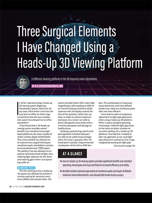
Three Surgical Elements I Have Changed Using a Heads-Up 3D Viewing Platform
RT Retina Today January/February 2019. A different viewing platform in the OR requires some adjustment. By S.K. Steven Houston III, MD.
In 2018, I operated using a heads-up 3D viewing system (Ngenuity Visualization System, Alcon) for all my cases. What better time than the new year to reflect on three surgical elements that this new visualization system has allowed me to refine and perfect?
I have found that a 3D heads-up viewing system provides several benefits over standard microscope-based platforms, the most notable of which is better depth of field (DoF). This enhanced DoF has allowed me to improve two surgical techniques: membrane peels and diabetic tractional retinal detachment (TRD) repair. The platform has also allowed me to operate with lower levels of light, thus reducing light exposure on the retina and reducing glare when I am operating under air.
MEMBRANE PEELS
The first technique that a heads-up 3D system has allowed me to hone is macular peeling for epiretinal membrane (ERM) and macular holes. The system provides better DoF under high magnification when peeling an ERM or an internal limiting membrane (ILM). I operate with the display toward the foot of the stretcher, rather than up close, in order to achieve maximum stereopsis. As a result, I am able to better distinguish tissue levels and to minimize disruption and damage to healthy tissue.
Initiating a peel during a pinch-and-peel approach is flawless because I am able to see subtle tissue changes when the tissue is grasped. After the initial peel is started, I enjoy enhanced visualization of the ILM or ERM borders. The combination of improved tissue distinction and more defined borders has made my membrane peels safer and more efficient.
I have had to make an important adjustment to light pipe placement when using a heads-up 3D platform. When I used a standard operating microscope, I held the light pipe close to the retina to better visualize tissue when peeling. On a heads-up 3D platform, I learned that I needed to alter my approach, as my usual light pipe placement washed out the image. I adapted by backing the light pipe away from the retina just slightly, which resulted in a crisp, perfect image.
DIABETIC TRDs
My technique to surgically repair diabetic TRDs has also improved with a heads-up 3D platform. On a standard microscope, I used a flat contact lens for high magnification viewing during surgery in the macula. In diabetic patients, membranes often extend past the arcades into the mid-periphery. Lens manipulations, aberrations from the edge of the lens, and small pupils can all contribute to limited viewing past the arcades with a flat macular contact lens and can quickly degrade the view.
On a heads-up 3D viewing platform, I perform all segmentation and delamination of membranes in diabetic patients with TRD with the BIOM lens (Oculus) under high magnification. I can even address membranes in the mid-periphery. The platform’s enhanced DoF allows me to maintain good focus despite the high magnification, decreasing my reliance on contact lens viewing. Just as the platform’s enhanced DoF has improved the safety and efficiency of ILM and ERM peels, so too has the platform made diabetic TRD cases safer and more efficient.
LIGHT LEVELS
I now use lower light levels during surgery. Lower levels of light pipe illumination can be used without a noticeable difference for standard maneuvers, thanks to the digitized viewing on this platform. The lower light levels possible with this system also help reduce the glare we encounter when using a standard operating microscope and working under air.
I have experimented with decreasing light pipe illumination while adjusting the digital viewing system and have found that light pipe illumination levels can be decreased from the 25-35 setting to 5 on the Constellation Vision System (Alcon) without compromising surgical viewing, significantly reducing retinal light exposure and enhancing patient safety.
MORE TO COME?
After my first year using this viewing platform full time, these three factors stick out in my mind as reasons that I’ve made a complete switch from analog to digital surgical viewing. I look forward to the continued development of 3D digital viewing. I plan on perfecting more techniques over the next year, and I am eager to see how my experience this year will shape my future surgical approaches.
 Click to read or print the article.
At a Glance
- Use of a heads-up 3D viewing system provides significant benefits over standard operating microscopes and may contribute to increased efficiency and safety.
- Benefits include improved approaches to membrane peels and repair of diabetic tractional retinal detachments, and reduced light levels during surgery.
 S.K. Steven Houston III, MD
Vitreoretinal Surgeon, Florida Retina Institute, Orlando, Florida
Editorial Advisory Board Member, Retina Today
Financial disclosure: None
Please click here to view the full page digital version of RT Retina Today January February http://retinatoday.com/2019/02/